MicroCT is a powerful tool that can be used to non-destructively generate high-resolution two- and three-dimensional (2D/3D) representations of an object’s internal and external structure down to the submillimeter, micro- (1–100 microns) and even nano-scales (< 1 micron). Check out our Equipment Page to learn more about the basics of microCT and the specific equipment at MICRO.
MicroCT is applicable to a wide range of disciplines including mechanical engineering, biology, anthropology, anatomy, paleontology, geology, neuroscience, chemistry and biochemistry, human health, biomedical engineering, and beyond.
Click on the images below to see some examples or feel free to contact us at micro@uark.edu if you are interested in finding out about potential applications of microCT for your own research.
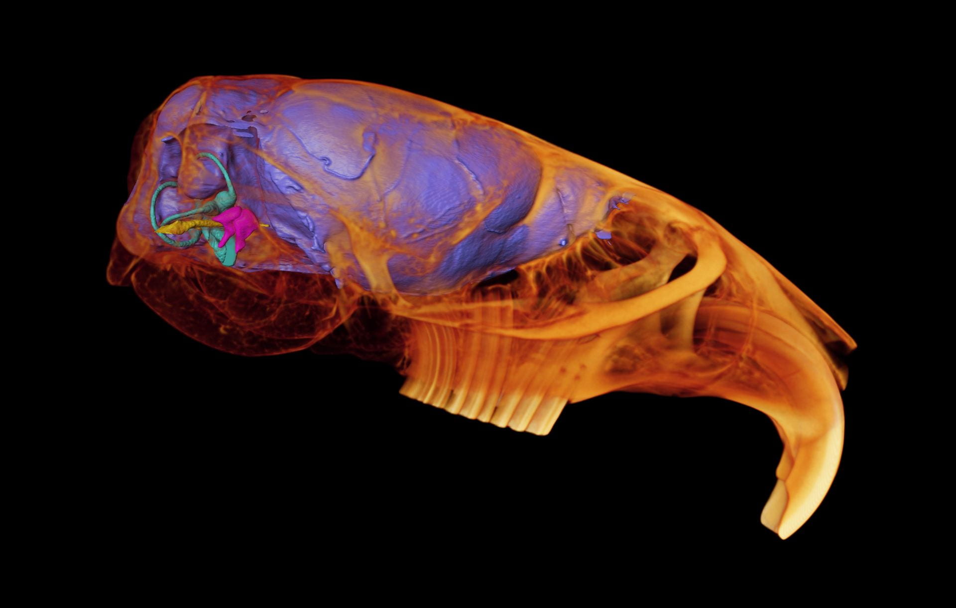
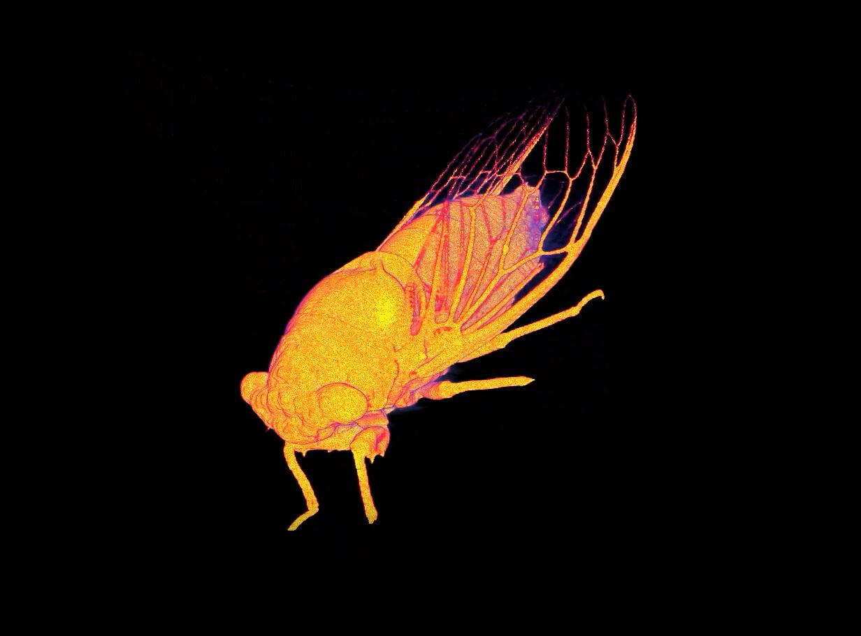
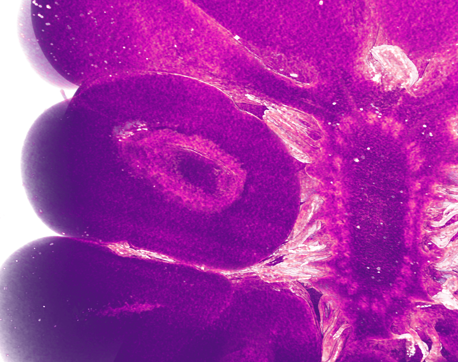
Zoological and anatomical specimens such as hearts, brains, snakes, birds, primates, insects, fish, rats, lizards, alligators, pigs, possums, goats, and many more


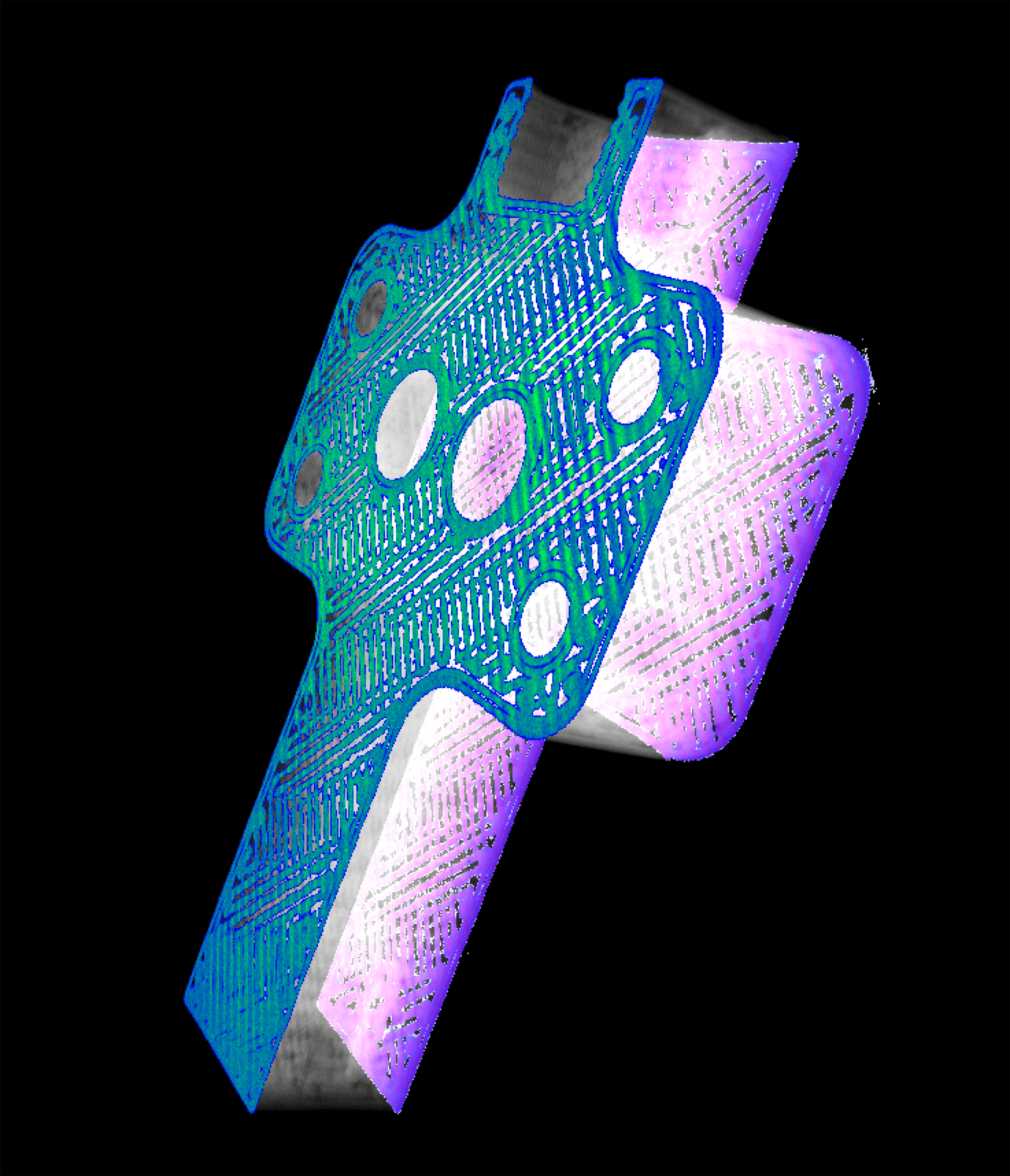
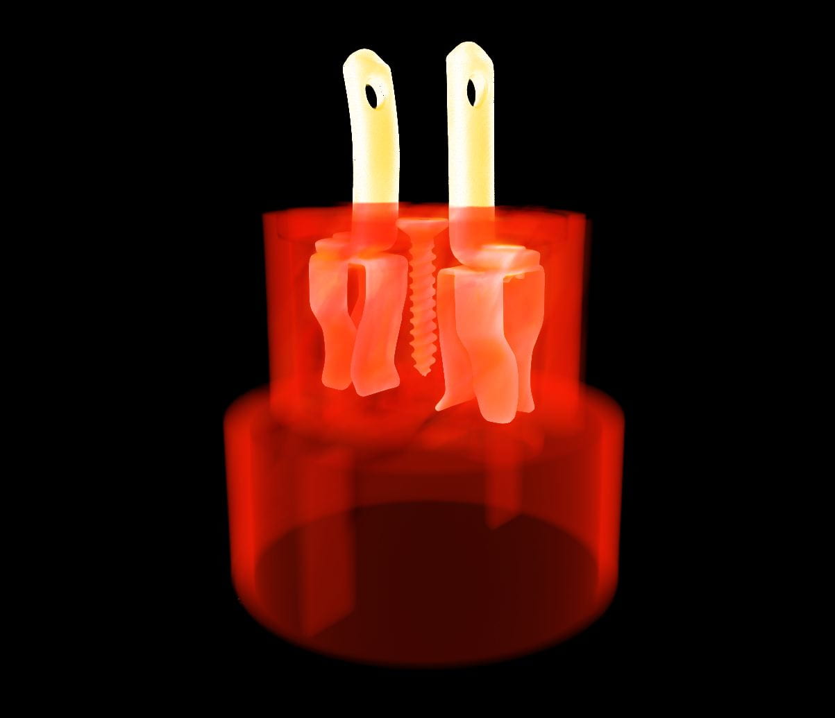
Manufactured objects such as computer parts, heart stents, cement, plastic parts, fused metal objects, or 3D printed metal

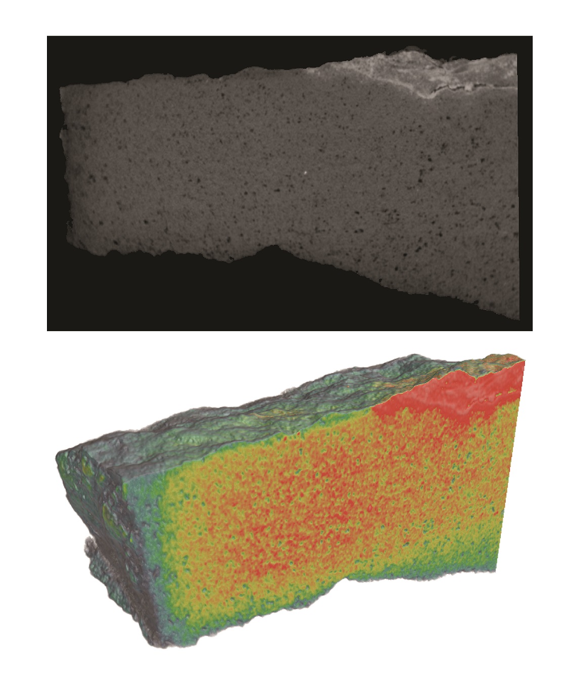
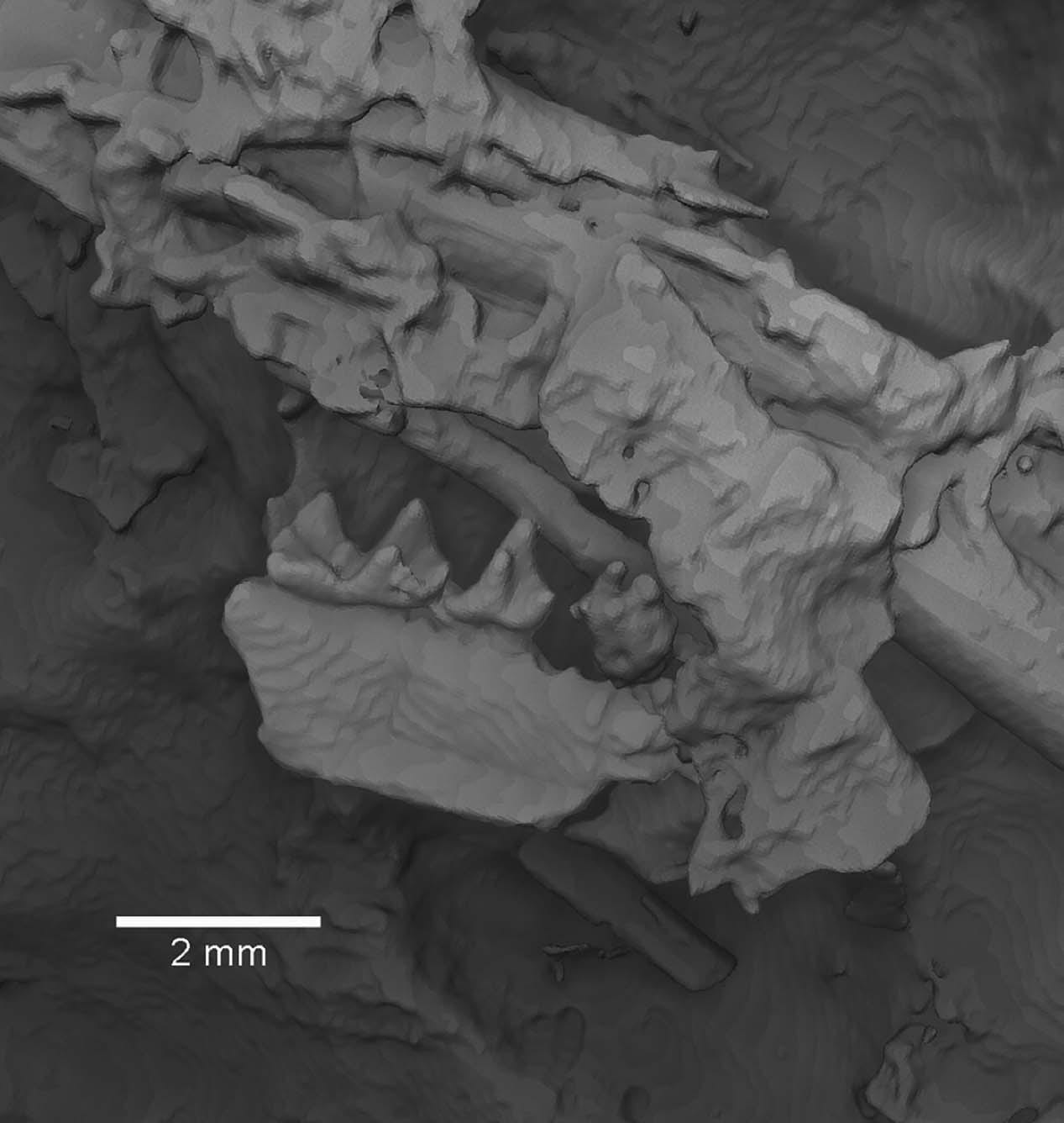
Geological and paleontological items such as rock samples and cores, as well as fossil remains

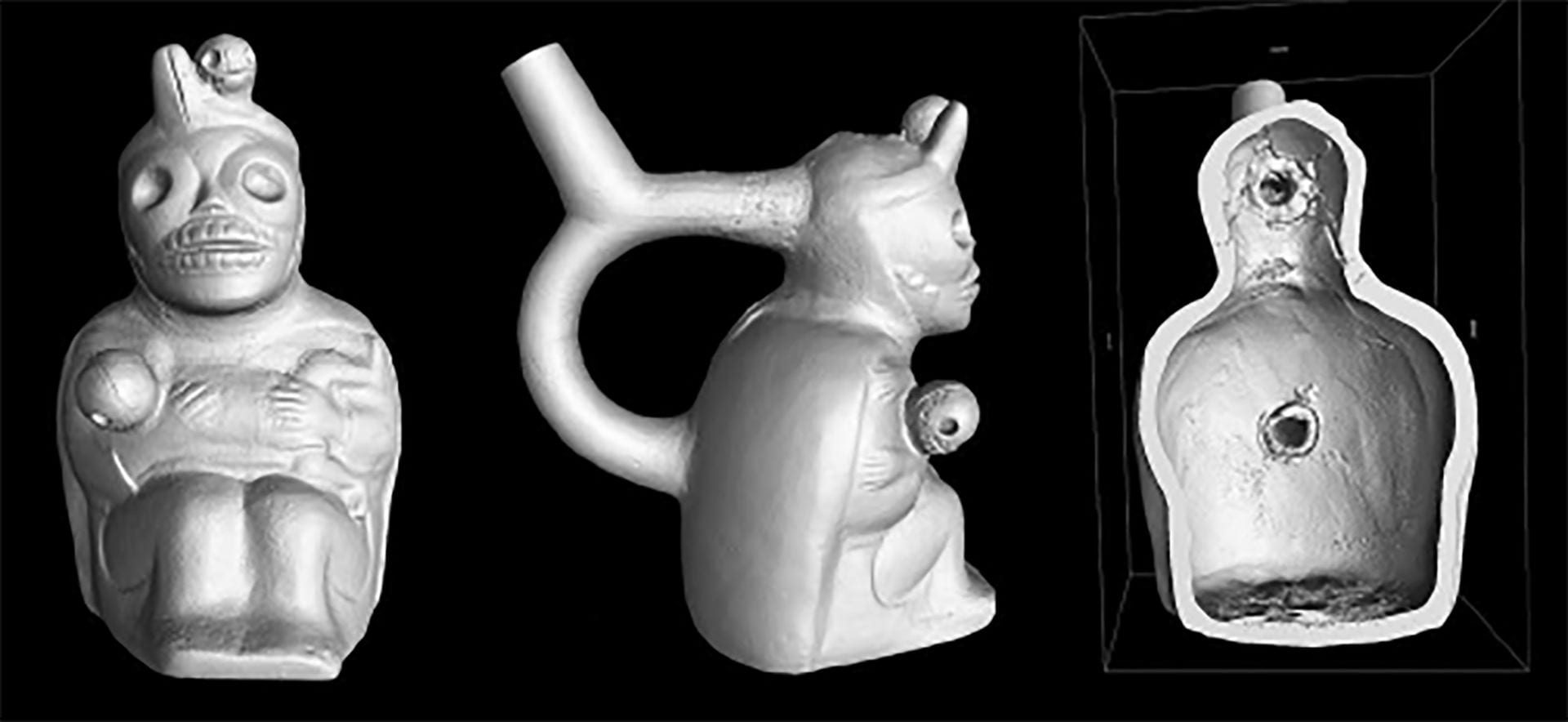
Archaeological objects including ceramic vessels, sherds, reed flutes, and seeds


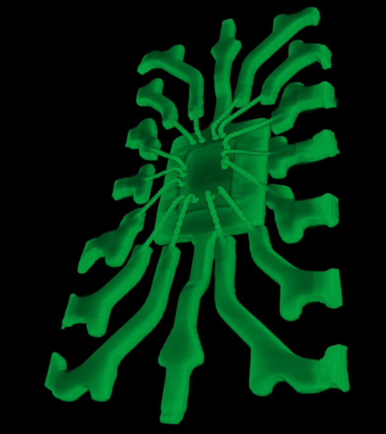
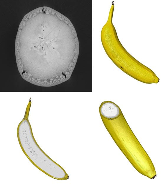
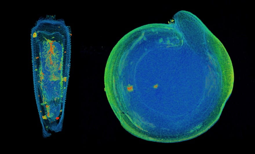
Botanical specimens such as fruits and vegetables, seeds, pinecones, and more

MICRO Publications
You can also find publications using MICRO data via Google Scholar
2025
- Lee A, Moore JM, Covarrubias BV, Lynch LM. 2025. Segmentation of cortical bone, trabecular bone, and medullary pores from micro-CT images using 2D and 3D deep learning models. The Anatomical Record.
- Taylor AB, Holmes MA, Laird MF, Terhune CE. 2025. Jaw-Muscle Structure and Function in Primates: Insights into Muscle Performance and Feeding-System Behaviors. Evolutionary Anthropology 34, e22053.
2024
- Forcellati MF, Green TL, Watanabe A. 2024. Brain shapes of large-bodied, flightless ratites (Aves: Palaeognathae) emerge through distinct developmental allometries. Royal Society Open Science 11, 240765.
- Gignac PM, Aceves V, Baker S, Barnes JJ, Bell J, Boyer D, Cunningham C, De Carlo F, Chase MH, Cohen KE, Colbert M, De Cree T, Daza J, Dickinson E, DeLeon V, Dougan L, Duffy F, Dunham C, Early C, Edey D, Echols S, Eckley SA, Fenner K, Franklin KP, Gila B, Goetz FB, Gray JA, Gleiber D, Hall AS, Hanna R, Hannula M, Harris W, Hill JJ, Holliday CM, Hurdle K, Jayarajan A, Knaub JL, Krause AR, Leavey A, Lessner EJ, Lynch LM, Maga M, Maisano J, Marsh K, Marsh M, Martin-Silverstone E, Misiaszek J-P, Neander AI, O’Brien HD, Olson S, Panigot E, Motch Perrine SM, Porri T, Ramsey A, Scheiffele G, Smith HF, Stanley EL, Stock SR, Terhune CE, Thomas DL, Vargas CAL, Veltri M, Warnett JM, Watanabe A, Waters EA, Wende R, Wescott DJ, Withnell CB, Whittaker S, Wilbur ZE, Wilson J, Wilson M, Winchester J, Yoakum CB, Zobek CM. 2024. The role of networks to overcome large-scale challenges in tomography: The non-clinical tomography users research network. Tomography of Materials and Structures 5, 100031.
- Holmes MA, Terhune CE, Chalk-Wilayto J, Yoakum CB, Taylor PM, Ramirez R, Solis M, Polvadore TA, Ross CF, Taylor AB, Dutra Fogaca M, Laird MF. Ontogenetic changes in jaw leverage and skull shape in tufted and untufted capuchins. Journal of Morphology 285(5), e21705.
- Kay D. 2024. Dental Alveoli and the Evolution of Complex Dentitions in Tetrapods: Patterns, Trajectories, and Implications. PhD Dissertation. Oklahoma State University Center for Health Sciences.
- Knoll, F, Ishikawa, A, Kawabe S. 2024. A proxy for brain-to-endocranial cavity index innon-neornithean dinosaurs and other extinct archosaurs. Journal of Comparative Neurology 532(3), e25597.
- Polvadore TA, Yoakum CB, Taylor PM, Holmes MA, Laird MF, Chalk-Wilayto J, Kanno CM, de Oliveira JA, Terhune CE. 2024. Ontogenetic biomechanics of tufted (Sapajus) and untufted (Cebus) capuchin mandibles. American Journal of Biological Anthropology 185(2), e25006.
- Straight PJ, Gignac PM, Kuenzel WJ. 2024. A histological and diceCT-derived 3D reconstruction of the avian visual thalamofugal pathway. Scientific Reports 14, 8447.
- Yoakum CB, and CE Terhune. 2023. The inferior alveolar nerve and its relationship to the mandibular canal. The Anatomical Record 307(1), 97-117.
2023
- Frosali S, Bartolini-Lucenti S, Madurell-Malapeira J, Urciuoli A, Costeur L, Rook L. 2023. First digital study of the frontal sinus of stem-Canini (Canidae, Carnivora): evolutionary and ecological insights throughout advanced diagnostic in paleobiology. Frontiers in Ecology and Evolution 11, 1173341.
- Green TL, and Gignac PM. Osteological comparison of casque ontogeny in palaeognathous and neognathus birds: insights for selecting modern analogues in the study of cranial ornaments from extinct archosaurs. Zoological Journal of the Linnaean society 199(1), 10-25.
- Klehm C. 2023. The use and challenges of spatial data in archaeology. Advances in Archaeological Practice 11 (1), 104-110.
- Plateau O, Green TL, Gignac PM, Foth C. 2023. Comparative digital reconstruction of Pica pica and Struthio camelus and their cranial suture ontogenies. The Anatomical Record 307(1), 5-48.
- Sender R. 2023. The biomechanics of tooth strength in Australopiths. PhD Dissertation, Washington University in St. Louis.
- Stephens S. 2023. Mitral valve imaging and biomechanics: A workflow towards computational modeling and validation. MA Thesis, University of Arkansas.
- Straight PJ, Gignac PM, Kuenzel WJ. 2023. Mapping the avian visual tectofugal pathway using 3D reconstruction. The Journal of Comparative Neurology 532(2), e25558.
- Watanabe A, Marshall SS, and PM Gignac. 2023. Dumbbell-shaped brains of Polish crested chickens as a model system for the evolution of novel brain morphologies. Journal of Anatomy 243(3), 421-430.
2022
- Cooper AR. 2022. Primate olfaction: a phylogenetic analysis of cribriform plate morphology. Undergraduate honors thesis, University of Arkansas.
- Croghan J. 2022. Assessment of Cranial Morphology and Function Underlying Dietary Diversity in Cryptodires. PhD Dissertation. Ohio State University.
- Czaplewski NJ, GS Morgan, RJ Emry, PM Gignac, and HD O’Brien. 2022. Three new Early-Middle Eocene bats (Mammalia: Chiroptera) from Elderberry Canyon, Nevada, USA. Smithsonian Contributions in Paleontology 106.
- George J. 2020. A reanalysis of the anole dewlap at the osteological, histological, and myological levels. MA Thesis, Oklahoma State University Center for Health Sciences.
- Green TL, DI Kay, and PM Gignac. 2022. Intraspecific variation and directional casque asymmetry in adult southern cassowaries (Casuarius casuarius). Journal of Anatomy 241, 951-965.
- Feldman KM, YA O’Keefe, PM Gignac, and HD O’Brien. 2022. Highest resolution microCT scan of the human brainstem reveals putative anatomical basis for infrequency of Medial Medullary Syndrome. NeuroImage: Clinical 36, 103272.
- Stephens SE, Ingels NB, Wenk JF, and MO Jensen. 2022. Alumina as a computer tomography soft material and tissue fiducial marker. Experimental Mechanics 62, 879-884.
- Straight P. 2022. Utilizing the 3D Environment to Facilitate Learning of Complex Visual Neural Pathways in the Avian Brain. MS Thesis, University of Arkansas.
2021
- Olson RA, SJ Montuelle, H Curtis, and SH Williams. 2021. Regional tongue deformations during chewing and drinking in the pig. Integrative Organismal Biology 3(1), obab012.
- Marbut C, B Nafis, and D Huitink. 2021. Accelerated mechanical low cycle fatigue in isothermal solder interconnects. Microelectronics Reliability 116, 113998ff.
- Stephens SE, M Bean, H Surber, NB Ingels, HK Jensen, S Liachenko, JF Wenk, and MO Jensen. 2021. MicroCT imaging of heart valve tissue in fluid. Experimental Mechanics 61, 253-261.
- Taylor MS. 2021. Assessing morphological changes to the periaqueductal gray and the corpus collosum in response to opioid exposure in an adolescent rat model. MA Thesis, Oklahoma State University Center for Health Sciences.
- Yoakum CB. 2021. An assessment of the neurovascular structure of the trigeminal nerve and their relationship to diet in primates. PhD Dissertation, University of Arkansas.
2020
- Farr CL. 2020. Permeability Anisotropy and Meandering Fluvial Facies Architecture of the Bartlesville Sandstone, Nowata County, Oklahoma. MA Thesis, University of Tulsa.
- Green TL. 2020. Ontogeny, disparity, and function of the enigmatic casques of cassowaries (Casuarius): A case study of cranial ornamentation in archosaurs. PhD Dissertation, Oklahoma State University Center for Health Sciences.
- Green TL, and PM Gignac. 2020. Osteological description of casque ontogeny in the southern cassovary (Casuarius casuarius) using micro-CT imaging. The Anatomical Record 304, 461-479.
- Olson RA. 2020. Biomechanics of the mammalian Tongue: Kinematic analysis of tongue movements and deformations during feeding and drinking. PhD Dissertation, Ohio University.
- Terhune CE, AD Sylvester, JE Scott, and MJ Ravosa. 2020. Internal architecture of the mandibular condyle of rabbits is related to dietary resistance during growth. Journal of Experimental Biology 223, jeb220988.
2019
- Plavcan JM, CV Ward, RF Kay, and FK Manthi. 2019. A diminutive Pliocene guenon rom Kanapoi, West Turkana, Kenya. Journal of Human Evolution 135, 102623.
- Tadros M. 2019. Designing an in vitro mitral valve mounting and testing system for microCT imaging. Undergraduate honors thesis, University of Arkansas.
Other References
- Gignac PM, Kley NJ. 2014. Iodine-enhanced micro-CT imaging: Methodological refinements for the study of soft-tissue anatomy of post-embryonic vertebrates. Journal of Experimental Zoology B 322B: 166–176.
- Gignac PM, Kley NJ. 2018. The utility of diceCT imaging for high-throughput comparative neuroanatomical studies. Brain, Behavior and Evolution 91: 180-190.
- O’Brien HD, Gignac PM, Hieronymus TL, Witmer LM. 2016. Post-natal growth of the cranial arteries of the giraffe (Artiodactyla: Giraffa camelopardalis). PeerJ 4:e1696.